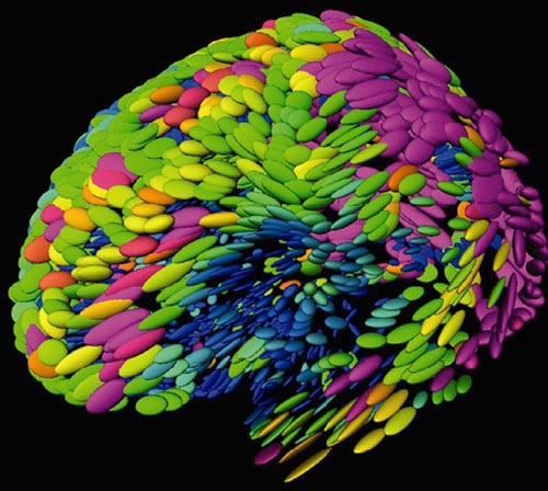
Using computer software programs, scientists combined brain MRIs from 20 normal people into this composite image, in which ellipsoids represent normal anatomical variations.


Using computer software programs, scientists combined brain MRIs from 20 normal people into this composite image, in which ellipsoids represent normal anatomical variations. Pink purple ellipsoids, signifying the greatest variation, occur in brain regions that are uniquely human for example, regions that control language and logical reasoning. Blue ellipsoids, representing slight variations, occur in brain regions that control sensation and movements. Ultimately, this baseline data on interpersonal variability will allow scientists to distinguish normal anatomical variation from abnormal brain loss, such as that seen in Alzheimer's disease.
Image courtesy of Dr. Paul Thompson, University of California, Los Angeles.
